
Understanding Eye Floaters: Causes, Symptoms, & Treatment Options
Probably the most common question I have in clinic is about floaters in the vision. But what exactly are they? I thought I’d write about this in the blog this month as last week I had a sudden, new, very large floater and it needed assessment. More about that later…
To understand floaters first you need to appreciate the anatomy of the eye:

Anatomy of the eye
The eye is bit like a ping pong ball – but the inside of the eye is filled with a jelly like substance called vitreous. Just like the air in ping pong ball the jelly cannot escape from the eye. When floaters occur, they are due to a change in this vitreous jelly. The vitreous can change for several reasons – the most serious is retinal tear or detachment. More usually the vitreous changes due to it becoming more liquid and it shifts about. The floaters cast little shadows on the retina that move with your vision. They can be extremely irritating, but the brain is pretty good as blocking them out over time.
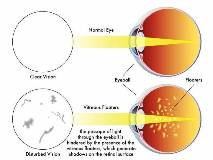

This is what the vitreous floaters can look like
The most important thing is getting any new floaters assessed. This is to rule out a retinal tear or detachment which is sight threatening. Other signs of retinal detachment can be loss of vision, shadows across vision and/or flashes of light. If you are in any doubt, please contact the clinic.
After my sudden new floater, I had an Optomap scan and this showed a large floater near the optic nerve – a retinal detachment was ruled out. Here’s my scan with the vitreous floater highlighted:
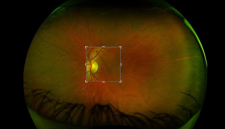
Optompap Retinal Scan
So, what can be done about floaters?
If your vitreous floaters get in the way of your vision, which happens rarely, you and may consider treatment. Options may include surgery to remove the vitreous or a laser to disrupt the floaters, although both procedures are rarely done. The NHS will not routinely fund this treatment so usually has to be performed at a private eye hospital by a vitreo-retinal surgeon.
Surgery to remove the vitreous: An ophthalmologist who is a specialist in retina and vitreous surgery removes the vitreous through a small incision (vitrectomy). The vitreous is replaced with a solution to help your eye maintain its shape. Surgery may not remove all the floaters, and new floaters can develop after surgery. Risks of a vitrectomy include infection, bleeding and retinal tears.
Using a laser to disrupt the floaters: An ophthalmologist aims a special laser at the floaters in the vitreous (vitreolysis). This may break up the floaters and make them less noticeable. Some people who have this treatment report improved vision; others notice little or no difference. Risks of laser therapy include damage to your retina if the laser is aimed incorrectly.
If you’d like to know more about floaters please follow this link. Advice taken from literature provided by The Association of Optometrists.
I look forward to welcoming you in practice again soon.
Lucinda
*The information presented here reflects general information about floaters but of course is not exhaustive
Have you read our other blogs?


-
2025
- 15 Apr 2025 Spring eye care: Protecting and prioritising your vision 15 Apr 2025
- 18 Mar 2025 Pro Tips for Proper Eyewear Care 18 Mar 2025
- 18 Feb 2025 Night Driving and Vision: Key Challenges and Solutions 18 Feb 2025
- 16 Jan 2025 Understanding different types of spectacle lenses 16 Jan 2025
-
2024
- 31 Oct 2024 Diabetic Eye Disease Month: The Power of Pre-Diabetes Checks 31 Oct 2024
- 19 Sept 2024 Exciting Developments in Ocular Science 19 Sept 2024
- 20 Aug 2024 The Benefits of UV Protection for Your Eyes 20 Aug 2024
- 1 Aug 2024 Tips for Maintaining Healthy Vision as You Age 1 Aug 2024
- 4 Jul 2024 The link between nutrition and eye health 4 Jul 2024
- 12 Jun 2024 How to Relieve Digital Eye Strain 12 Jun 2024
- 16 May 2024 Exploring Menopause's Impact on Eye Health 16 May 2024
- 18 Apr 2024 The Importance of Regular Eye Exams for Optimal Vision Health 18 Apr 2024
-
2023
- 14 Dec 2023 Understanding Macular Degeneration: A Guide to Age-Related Vision Changes 14 Dec 2023
- 21 Nov 2023 Adaptive Vision: Unveiling the Wonders of Photochromic Lenses 21 Nov 2023
- 26 Oct 2023 Transform Your Look Safely with Costume Contact Lenses! 26 Oct 2023
- 28 Sept 2023 Cutler and Gross - 50 years of style 28 Sept 2023
- 8 Aug 2023 AI Revolutionizing Optometry Care 8 Aug 2023
- 11 Jul 2023 Unlocking the Power of Optomap Scanning 11 Jul 2023
- 6 Jun 2023 Discover the Benefits of Polarized Sunglasses 6 Jun 2023
- 11 May 2023 Understanding Dry Eye (Keratoconjunctivitis Sicca) 11 May 2023
- 25 Apr 2023 Case Study: Understanding the Impact of a Retinal Tear 25 Apr 2023
- 21 Mar 2023 Ocular Hypertension: A Case Study 21 Mar 2023
- 28 Feb 2023 Unveiling the Process of Cataract Formation 28 Feb 2023
- 4 Jan 2023 Case Study: TIA (Mini-Stroke) 4 Jan 2023
-
2022
- 21 Dec 2022 Tips for Winter Eye Care 21 Dec 2022
- 24 Nov 2022 Advice for soft contact lens wear 24 Nov 2022
- 25 Oct 2022 Understanding Eye Floaters: Causes, Symptoms, & Treatment Options 25 Oct 2022
- 22 Sept 2022 Hormonal Impact on Eye Health during Menopause 22 Sept 2022
- 16 Aug 2022 Arcus Senilis (corneal arcus) – What is it? 16 Aug 2022
- 28 Jul 2022 Light Sensitivity (Photophobia): Understanding Causes and Finding Relief 28 Jul 2022
- 23 Jun 2022 Why do you need longer arms as you grow older? 23 Jun 2022
- 19 May 2022 Best Practices for Contact Lens Wear & Care 19 May 2022
- 19 May 2022 Dive Into Clarity: Discover Prescription Swimming Goggles 19 May 2022
- 21 Apr 2022 The Next Exciting Step in Eye Care - Optomap 21 Apr 2022
- 15 Feb 2022 Unraveling the mystery of human crying 15 Feb 2022
-
2021
- 2 Dec 2021 Ready Readers vs Prescription Lenses: A Comprehensive Guide 2 Dec 2021
- 2 Nov 2021 How to clean your micro fibre lens cloth 2 Nov 2021
- 5 Oct 2021 8 Tips for Protecting Your Eyes & Keeping Them Healthy 5 Oct 2021
- 7 Sept 2021 Unlocking the Mysteries of Astigmatism 7 Sept 2021
- 20 Jul 2021 Cataract: Unveiling the facts, symptoms & treatment 20 Jul 2021
- 3 Jun 2021 OCT as standard on all eye tests 3 Jun 2021
- 14 May 2021 Unlocking Clarity: Strategies to Prevent Myopia Progression 14 May 2021
- 1 Apr 2021 Understanding the science of Blue Light-Blocking Lenses 1 Apr 2021
- 22 Mar 2021 Embrace the Brighter Days: Celebrating an Iconic British Brand This March 22 Mar 2021
- 22 Feb 2021 Combat Dry Eye and Allergies: Expert Solutions for Relief and Comfort 22 Feb 2021
- 21 Jan 2021 What Is Screen Fatigue? 21 Jan 2021
-
2020
- 22 Dec 2020 Polarised Lenses – The Most Effective Way to Reduce UV Radiation 22 Dec 2020
- 24 Nov 2020 Protect Against Dry Eyes in Cold Weather 24 Nov 2020
- 19 Oct 2020 DVLA Driving Standards – The Clear Facts 19 Oct 2020
- 16 Sept 2020 The Delight of Multifocal Contact Lenses 16 Sept 2020
- 15 Jul 2020 Night Driving – Can anything help to reduce glare? 15 Jul 2020